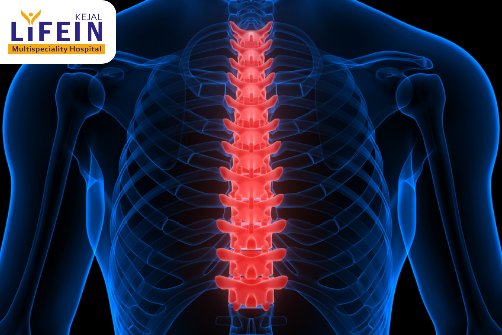
X-ray is a quick, painless procedure that is performed to create images of the structures within the body, particularly the bones. The bone appears as a white mass on the x-ray plate, whereas muscles and fat tissue appear as shades of grey.
X-rays are a type of radiation called electromagnetic waves. X-ray imaging creates pictures of the inside of your body. The images show the parts of your body in different shades of black and white. This is because different tissues absorb different amounts of radiation. Calcium in bones absorbs x-rays the most, so bones look white. Fat and other soft tissues absorb less and look gray. Air absorbs the least, so lungs look black.
The most familiar use of x-rays is checking for fractures (broken bones), but x-rays are also used in other ways. For example, chest x-rays can spot pneumonia. Mammograms use x-rays to look for breast cancer.
When you have an x-ray, you may wear a lead apron to protect certain parts of your body. The amount of radiation you get from an x-ray is small. For example, a chest x-ray gives out a radiation dose similar to the amount of radiation you’re naturally exposed to from the environment over 10 days.